No one knows a child's heart like SickKids
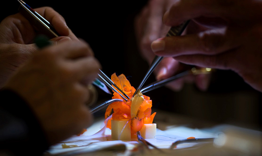
We teach surgeons to fix hearts by making them.3-D printing is here. Yes, you can now make a 3-D bobblehead of yourself. But SickKids cardiac radiologist Dr. Shi-Joon Yoo, and MRI technologist Omar Thabit, had a more revolutionary, and life-saving, application in mind for the technology. They decided to 'print' babies' hearts, using diagnostic imaging as their blueprint, to give surgeons a new learning tool. At first, the hearts were hard plastic. Although each still takes a day to print, they're now soft. And while not quite the same as real tissue, they're an effective medium in which surgeons can see, and operate on, a variety of congenital heart defects.
When 'practice' surgery is performed on a printed heart, "If there's a problem, no problem," says Dr. Glen Van Arsdell, Head of Cardiovascular Surgery at SickKids, who, with Dr. Yoo, recently welcomed 11 surgeons from across Canada - and around the globe, from Chile to Norway - to SickKids for what they call 'HOST': Hands-On Surgical Training.
This kind of training is very new – they've been doing it less than a year – and it's a world first.
The detail of the hearts makes them profoundly realistic. As Dr. Yoo says, "The highest resolution current medical imaging can give surgeons is 0.3 - 0.7 mm. The printer we use delivers 0.3 mm resolution." At a recent training session in SickKids Peter Gilgan Centre for Research and Learning, one of the visiting surgeons put it well: "Usually, I have to assemble a picture of the heart in my mind from a series of 2-D images. This way, I can see it."
That's a key benefit 3-D hearts offer to surgeons. But 3-D hearts will have a huge impact on patients and families, too. Traditionally, the discovery process happened in the operating room, with the child's chest open – creating the possibility of the negative impacts associated with major surgery on tiny children. Now, the operation can be rehearsed, and assistants can be as familiar with the defect as the primary surgeon. Rehearsal will save surgical time – and therefore time under anaesthesia – which both Dr. Yoo and Dr. Van Arsdell know leads to better outcomes.
In the recent training session Drs. Van Arsdell and Yoo 'HOST'ed, the surgeons worked with 5 models, representing a suite of 5 congenital heart defects. This initiative reinforces SickKids' commitment to training, and the hospital's well-earned reputation as a world leader in congenital heart disease. Drs. Van Arsdell and Yoo look forward to making this training standard for SickKids fellows – and for the hospital being seen, worldwide, as "the place to train."
